
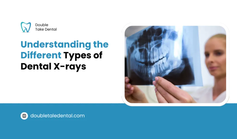
Cavities cannot be diagnosed or seen in detail with the naked eye. If dentists diagnose cavities using mirror tools, they need more advanced technology to determine the depth of the cavities. For this purpose, X-rays are used.
Dental X-rays can give an in-depth image of the cavities and help dentists to understand the severity of the problem.
Additionally, they allow dentists to detect and treat dental issues before they become serious and more painful. There are several types of dental X-rays. These different types of X-rays work for several reasons.
In this guide, we will explore these types and their functions in detecting cavities and dental infections. Let’s get started!
What Are Dental X-rays?
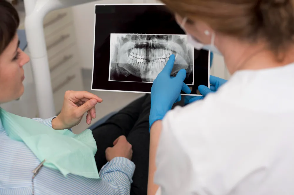
Dental X-rays are diagnostic imaging tools, similar to other diagnostic imaging methods used to examine the body. They capture pictures of your teeth, bones, and surrounding tissues. These images help dentists conduct an in-depth examination of tooth decay, any dental infections and bone loss that cannot be seen during visual exams.
There are two main types of dental X-rays:
- Intraoral X-rays (taken inside your mouth)
- Extraoral X-rays (taken outside your mouth)
Besides these X-rays, dentists are now using digital dental X-rays that use less radiation than the conventional types. Their reduced radiation exposure makes them a safer choice for regular use.
What Are Intraoral X-rays?
Intraoral X-rays are the most common type of dental X-rays. These are taken from inside your mouth and help your dentist get a clear look at your teeth, jawbone, and surrounding structures.
They are especially useful for detecting cavities, checking bone health, and examining individual teeth in detail.
Here are some types of intraoral X rays.
Bitewing X-rays
Bitewing X-rays are the most frequently performed X-rays during routine dental check-ups. They provide a comprehensive view of both upper and lower molars and bicuspids. Dentists utilize these X-rays to:
- Identify cavities between teeth
- Identify early signs of bone loss from gum disease
- Check the fit of crowns and the condition of fillings
Periapical X-rays
A dental periapical Xray is used to take a complete picture of a single tooth, including its surrounding bones, its crown, and the roots. This X-ray is specifically utilized for:
- Tooth abscesses
- Root infections
- Bone loss
- Fractures or trauma
This periapical radiographic image allows your dentist to detect deep-seated issues that could compromise oral health.
Occlusal X-rays
Occlusal X-rays are used to view the complete arch of the teeth, whether it is of the lower jaw or the upper jaw. It is used to diagnose:
- Jaw fractures
- Impacted teeth
- Cysts and tumors
- Tooth development in children
Pediatric dentists often rely on these X-rays to evaluate children’s oral growth.
What Are Extraoral X-rays?
Unlike intraoral X-rays, extraoral X-rays are taken from outside the mouth. They give your dentist a broader view of your jaw, skull, and overall oral structure. These are particularly useful for treatment planning and evaluating jaw-related issues. Here are the main types:
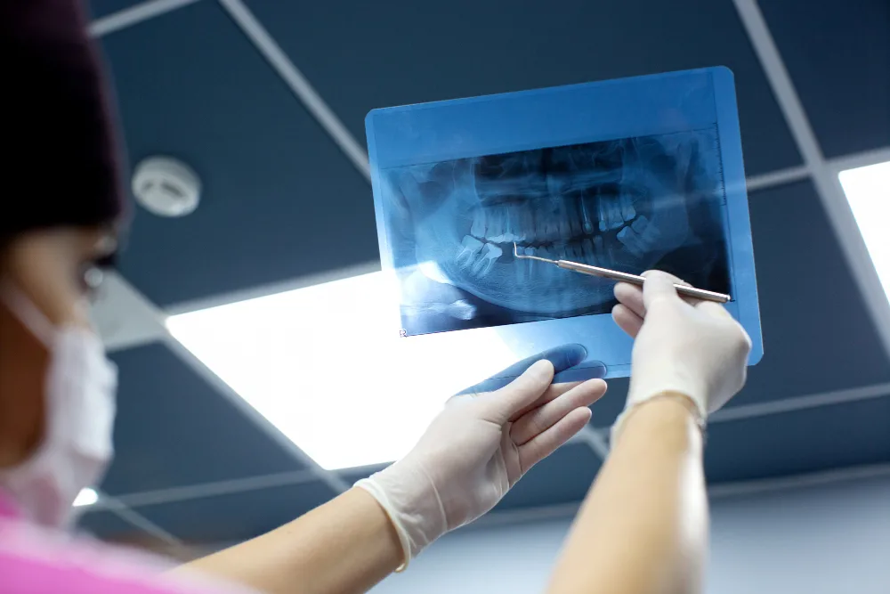
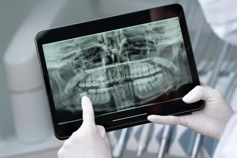
Panoramic X-rays
A panoramic X-ray provides a wide-angle view of your mouth in one image. It encompasses the upper and lower jaws, the sinus regions, and the temporomandibular joints (TMJ). Dentists typically use panoramic X-rays to:
- Check for impacted or emerging teeth
- Assess jaw structure
- Plan orthodontic treatments
Full Mouth X-rays
A full mouth Xray is a series of individual images, typically combining bitewing and periapical dental X rays. This detailed collection aids in identifying common dental problems and is frequently suggested for:
- New patient assessments
- Advanced gum disease
- Extensive dental work
Cephalometric and Cone Beam CT Scans
Advanced imaging like cephalometric X-rays and Cone Beam Computed Tomography (CBCT) provides 3D or lateral views of the skull, jaw, and teeth. These are typically used for:
- Orthodontic planning
- Dental implant placement
- Evaluating jaw disorders or abnormalities
These tools deliver high-resolution data that helps customize treatment plans with precision.
Comparison of the Most Common Dental X-ray Types
Out of these types of X-rays, some types are very similar to each other. Let’s compare a few in tabular form to know their differences.
Periapical vs. Bitewing
| Feature | Periapical X-ray | Bitewing X-ray |
| Image Scope | Entire tooth (crown to root) | Crowns of upper & lower back teeth |
| Best For | Root infections, trauma, abscesses | Cavities, bone loss, fillings |
| Coverage | One or two teeth per image | Multiple teeth per image |
Panoramic vs. Full Mouth
| Aspect | Panoramic X-ray | Full Mouth X-ray |
| Purpose | Provides a broad overview of oral structure | Provides detailed, close-up views of each tooth and supporting bone |
| Use Case | Recommended when a general overview is needed | Recommended when detailed information of each tooth is required |
| Imaging Coverage | Captures the entire mouth in one image | Multiple images covering all teeth and the surrounding bone |
Are Dentbal X-rays Safe?
Indeed, dental X-rays are safe when performed correctly. Using protective gear, such as lead aprons and thyroid collars, minimizes radiation exposure even more.
Digital X-rays result in lower radiation exposure compared to conventional film X-rays. A single digital bitewing X-ray exposes you to roughly the same amount of radiation as you naturally receive from your environment in one day.
That’s why they are considered a safer option as compared to traditional X-rays.
How Often Should You Get Dental X-rays?
The frequency of dental X-rays varies based on your age, oral health, and risk factors. General guidelines include:
- Children & teens: Every 6–12 months if there is a high risk of decay
- Adults with good oral health: Every 24–36 months
- Patients with gum disease or high decay risk: More frequent X-rays may be needed
Your dentist will establish your X-ray schedule according to your specific needs.
All-in-All
Dental X-rays are essential for diagnosing issues early, before they escalate into more severe problems. They help monitor deep cavities and infections that are not visible to the naked eye. However, various dental X-ray types are used for different purposes.
Intraoral x-rays are images of your jaws, bones, and muscles from inside the mouth. Extraoral x-rays are captured externally to provide a broader perspective of the skull and jaws. By learning about each type of X-ray and discussing it with your dentist, you can make a well-informed decision.
If you have any dental problems and are looking for a reliable solution, visit Double Take Dental. We offer efficient diagnosis and every type of solution with expert care. Our years of experience make us trustworthy to thousands of people.